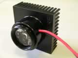
Design of condenser optics for the LED illuminator
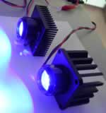
Using 28mm extension tubes from Comar Instruments, Cambridge, a 25mm diameter planoconvex lens with a focal length of 10mm (number) was mounted to collimate light from a 5W Royal Blue Lumiled. The Comar extension tubes are cheap and allow the direct internal mounting of standard 25mm diamter optics without the need for expensive adapters.
The collimated light from the LED is passed through a short pass filter, followed by a planoconvex lens with a focal length of 100mm, in order to focus the blue light at the back focal plane of the objective.
Beamsplitter and optical filters
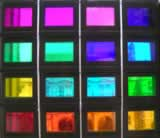
A filter block for a commercial fluorescence microscope typically costs around £400-£700. This is due to the use of expensive interference filters and dichroic beamsplitters. With the invaluable help of Dr. Brad Amos, MRC Laboratory of Molecular Biology, Cambridge, I have constructed a low cost alternative that is very effective for the detection of green fluorescent protein signals using blue LED illumination. Brad suggested that the beamsplitter could be made of Dichrolite, which are coated glasses produced for applications such as theatrical lighting. A variety of these glasses are available with different colours, and different dichroic properties. An image of a sampler of various dichrolite filters is shown below.
Using 28mm extension tubes from Comar Instruments, Cambridge, a 25mm diameter planoconvex lens with a focal length of 10mm (number) was mounted to collimate light from a 5W Royal Blue Lumiled. The Comar extension tubes are cheap and allow the direct internal mounting of standard 25mm diamter optics without the need for expensive adapters.
The collimated light from the LED is passed through a short pass filter, followed by a planoconvex lens with a focal length of 100mm, in order to focus the blue light at the back focal plane of the objective.
Beamsplitter and optical filters
A filter block for a commercial fluorescence microscope typically costs around £400-£700. This is due to the use of expensive interference filters and dichroic beamsplitters. With the invaluable help of Dr. Brad Amos, MRC Laboratory of Molecular Biology, Cambridge, I have constructed a low cost alternative that is very effective for the detection of green fluorescent protein signals using blue LED illumination. Brad suggested that the beamsplitter could be made of Dichrolite, which are coated glasses produced for applications such as theatrical lighting. A variety of these glasses are available with different colours, and different dichroic properties. An image of a sampler of various dichrolite filters is shown below.
Cheapo filter set for GFP fluorescence.
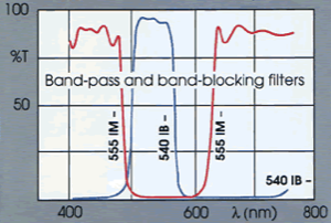
Excitation filter
555 IM 25
Magenta band blocking filter used as a short pass filter with a blue LED light source.
(Comar, £18)
555 IM 25
Magenta band blocking filter used as a short pass filter with a blue LED light source.
(Comar, £18)
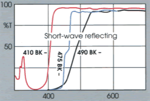
Dichroic beamsplitter
Dichrolite Y-54
Blue-reflecting mirror, mounted at 45 degrees to the light path.
(Comar, £25)
Dichrolite Y-54
Blue-reflecting mirror, mounted at 45 degrees to the light path.
(Comar, £25)
Emission filter
515 GY 25
Yellow Schott glass filter OG515, long pass filter.
(Comar, £14)
515 GY 25
Yellow Schott glass filter OG515, long pass filter.
(Comar, £14)
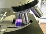
The dichroic filter is mounted at 45 degrees to the light path in a mirror block with C-mount threads (Edmund Optics). A 28mm to C-mount thread adapter (Comar) is used to mount the LED illuminator to the block. A C-T mount adapter is attached at the base of the filter block, and this clamps into the dovetail adapter of the Nikon Optiphot microscope shown here. T-mount components are 48mm in diameter and the microscope normally accepts a 49mm dovetail. A 25mm yellow Schott glass filter is fitted above the filter block to remove any reflected excitation light, and a C-mount video camera was used as a detector. I have experimented with a variety of low cost surveillance cameras that contain sensitive Sony ExView HAD CCDs. Video signals were passed to a video-firewire converter, and captured using a laptop computer with a firewire port
With a video camera placed on the filter block, all specimen positioning and focussing is done using the video output, displayed on a monitor or computer screen. Most firewire capture software provides a preview that is adequate for this. However, for lower cost and ease of use, direct inspection of fluorescence though the microscope would be a useful option. So I have experimented with the placement of an eyepiece above the filter block in this 160mm tube length microscope, and this works although it is a clumsy arrangement for viewing. The illuminator and filter block is now being adapted as an intermediate module for a microscope with infinity corrected optics.
A key feature of the cheaposcope design is the use of low cost optics and filters. For example, I have used a cheap colour process filter as the excitation filter. This is a filter designed for isolating the magenta component during CMYK imaging, and acts as a short pass filter when used with a blue light source. The beamsplitter is a cheap dicrolite filter, and the emission filter is alos a low cost longpass filter